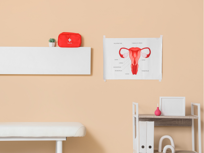
DEFINITION:
Hysteroscopy is the visualization of the entire uterine cavity using a hysteroscope, which is a fiber-optic instrument connected to a camera and covered by a metallic sheath. This sheath allows inflow and outflow of fluids for distension of the uterine cavity, in addition to an operative channel for introducing scissors, graspers, resectoscopes, and other instruments.
Hysteroscopy can be diagnostic, allowing direct visualization of the uterine cavity to detect abnormalities, and/or operative, enabling treatment of intrauterine pathologies such as polypectomy, fibroid resection, or septal removal.
Historical Background:
Hysteroscopy was first described by Bozzini in 1805 but was first used in gynecology by Pantaleoni in 1869 using a modified cystoscope. Further modifications were made to distend the uterine cavity using carbon dioxide in 1925 by Rubin. Later, distension by liquid media such as normal saline and 1.5% glycine by Gauss in 1934 replaced gas.
Continuous improvements in optics and illumination followed until 1970, when Hamou introduced micro-hysteroscopy and contact hysteroscopy. By 1980, operative hysteroscopy was introduced into clinical practice to manage various intrauterine pathologies.In the 1990s, office hysteroscopy became widely practiced.
From 2000 to the present, mini-hysteroscopes, vaginoscopy, and the no-touch technique have been introduced into modern practice.
In modern gynecology, diagnostic and operative hysteroscopy has become the gold standard for intrauterine evaluation and treatment of intrauterine lesions.⸻
Why and When to Use Hysteroscopy:
1. To correct myometrial abnormalities such as bicornuate uterus, unicornuate uterus, T-shaped uterus, uterine septum, and submucous fibroids.
2. To diagnose and treat endometrial disorders such as abnormal uterine bleeding, endometrial hyperplasia, atrophic endometrium, endometrial polyps, endometrial carcinoma, and thickened endometrium in postmenopausal women.
3. Family planning, including sterilization by blocking the proximal tubal ostium.
4. Exploration of the uterine cavity for foreign bodies such as a missed intrauterine device (IUD), removal of intrauterine bands, adhesions, and synechiae.
5. Excision of cervical polyps, resection of uterine septa involving the cervix, and dilatation of cervical stenosis.
6. In cases of recurrent IVF failure or recurrent pregnancy loss, and as part of routine infertility workup prior to IVF, such as excision of small polyps near the tubal ostium or facilitating difficult embryo transfer due to cervical stenosis.
7. Embryoscopy.
8. As a complementary procedure with laparoscopy for correction of cesarean section niche.
Contraindications of Hysteroscopy:
1. Pregnancy.
2. Acute pelvic inflammatory disease (PID).
3. Heavy uterine bleeding.
4. Cervical malignancy.
Setting:
1. Establishing the diagnosis using pelvic transvaginal ultrasound, hysterosonography, 3D ultrasound, and sometimes pelvic MRI with contrast for better imaging.
2. Explanation of the procedure to the woman prior to intervention and obtaining informed consent.
3. You can perform Hysteroscopy as an outpatient procedure and is usually well tolerated, especially with office hysteroscopy using local anesthesia via paracervical or intracervical injection. If the procedure remains painful or poorly tolerated, offer nitrous oxide mask. Oral NSAIDs such as ibuprofen (Brufen) prior to the procedure are routine and considered good practice for pain relief.
Hysteroscopy under general anesthesia in the operating theater is another option, depending on surgeon experience, patient cooperation, and the indication for hysteroscopy, whether diagnostic or operative.
Instruments Used During Operative Hysteroscopy:
1. Mechanical instruments:
• Scissors: Used for cutting adhesions or small polyps. They are slow and may cause some bleeding but result in no thermal damage.
• Graspers: Used for removal of polyps and foreign bodies such as IUDs.
2. Electrosurgical instruments:
• Resection loop (monopolar, bipolar, or true bipolar): Cuts tissue in slices and is effective for large lesions such as polyps, submucous fibroids, and endometrial resection. However, it carries risks of fluid overload , thermal injury and requires surgical expertise.
• Roller ball: An electrosurgical tool that uses electrical energy to coagulate the endometrium.
3. Mechanical morcellator systems:
Morcellators use rotating blades or shavers to cut and aspirate tissue imultaneously. They are indicated for endometrial polyps and small to moderate submucous fibroids. They work faster, cause less bleeding, provide clearer visualization, result in less fluid absorption, and eliminate the risk of electrical injury.
4. Laser hysteroscopy needs operator high experience.
Complications:
Complications of hysteroscopy are uncommon in experienced hands. However, the risk of uterine perforation increases in cases of cervical stenosis, such as in nulliparous and postmenopausal women, which may lead to false passage during hysteroscope insertion. Uterine perforation may be associated with urinary bladder or intestinal injury.
The surgeon must carefully monitor the volume of distension fluid used by calculating fluid inflow and outflow to avoid excessive fluid deficit, which can result in fluid overload, hyponatremia, pulmonary edema, and gas embolism.
Bleeding is another possible complication, usually due to excessive resection or myometrial injury. Such cases may require balloon tamponade and can progress to laparotomy.
Energy-related complications include thermal and electrical injuries or burns.
Infection, such as endometritis or pelvic infection may occur and can lead to intrauterine adhesions and subfertility, in addition to general anesthesia-related complications.
Conclusion:
Modern minimally invasive surgery such as hysteroscopy is now widely practiced worldwide, offering optimal diagnostic and operative interventions with minimal complications. It reduces hospital stay, minimizes pain, and allows rapid recovery.
We owe much to modern advances in optical technology and artificial intelligence, which have made full visualization of the uterine cavity a clinical reality.
Dr. Asmaa Abdulsalam
Consultant of Obstetrics and Gynecology
Al Ahli Hospital
Doha – Qatar


